

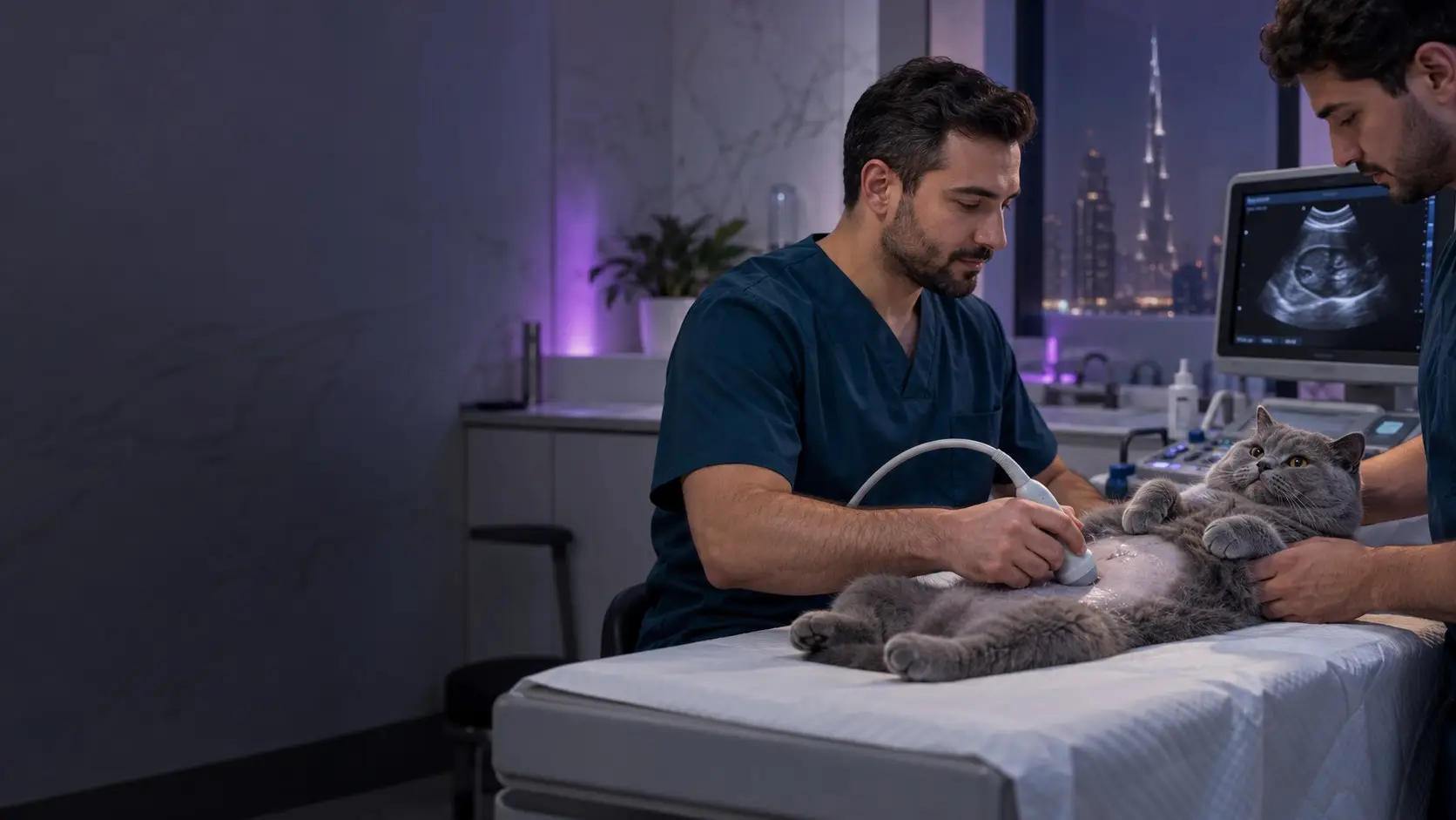
X-rays show bone, the chest and anything that holds gas or mineral. Ultrasound shows the inside of soft organs and any fluid around them. Your vet picks the one that answers the question, and in a sick animal with a belly problem that is often both.
The two tests do not compete. An x-ray is a single still picture of the whole area, made of shadows. Ultrasound is a moving picture of one small slice at a time. Each is blind to things the other sees clearly.
What an x-ray shows well
- Bones and joints. Fractures, dislocations, hip and elbow shape, arthritis, bone tumours.
- The chest. Lung patterns, fluid or air around the lungs, the size and outline of the heart, the windpipe.
- Gas patterns in the gut. A blocked intestine traps gas in a way that stands out. A twisted stomach in a large dog is diagnosed on one x-ray.
- Mineral and metal. Most bladder stones, swallowed coins, needles, stones and bone fragments.
- Late pregnancy. Once the skeletons have mineralised, puppies and kittens can be counted.
The weakness is soft tissue. On an x-ray the liver, spleen, fluid and a tumour are all the same shade of grey. A belly full of fluid becomes one blank area with no detail.
What ultrasound shows well
- The inside of organs. Liver texture, kidney structure, cysts, the layers of the gut wall, the spleen, lymph nodes.
- The bladder and womb. Wall thickness, sludge, small stones, polyps, and a fluid-filled womb in an unspayed female.
- Free fluid. A few millilitres of blood or urine in the abdomen show up, and the vet can guide a needle to sample them.
- Movement. Gut contractions, a beating foetal heart, blood flow.
- Some foreign objects. Cloth, rubber and fruit stones often cast no clear x-ray shadow but can be found on a scan.
The weakness is air and bone. Sound waves bounce off both. Ultrasound cannot see into normal lungs or through a gas-filled stomach, and it shows only the surface of a bone. It also depends heavily on the skill and patience of the person holding the probe.
Which test for which problem
| Problem | First test | Often added |
|---|---|---|
| Limping or a suspected fracture | X-ray | None in most cases |
| Coughing or laboured breathing | Chest x-ray | Heart scan if the heart looks enlarged |
| Repeated vomiting, possible swallowed object | X-ray | Ultrasound when the x-ray is unclear |
| Straining to urinate or blood in urine | Ultrasound or x-ray | The other one, plus a urine test |
| Raised kidney or liver values on bloods | Ultrasound | X-ray seldom needed |
| Swollen belly | Ultrasound | X-ray of chest and abdomen |
| Fall from height or road accident | Chest and limb x-rays | Quick ultrasound check for internal bleeding |
| Pregnancy | Ultrasound early | X-ray late, to count |
Three Dubai cases where both are used
The cat that fell from a balcony
Cats that fall from apartment balconies and windows often land on their feet and walk away. Many still have a bruised or collapsed lung, a split palate or a cracked pelvis. Chest x-rays come first, because a lung injury changes how safe sedation is. A short ultrasound sweep then checks the bladder and looks for free blood in the abdomen.
The dog that swallowed a date stone
Dates are in most homes here, and at every table in Ramadan. A date stone is plant material and may not show on an x-ray the way a pebble or bone does. The x-ray can still reveal the gas pattern of a blockage.
Ultrasound can then find the object itself and show whether the gut around it is still moving. Our page on swallowed foreign objects explains what follows.
The cat straining in the litter tray
Concentrated urine in a hot climate favours crystals and bladder stones. Most stones are visible on x-ray, which also shows stones lodged in the urethra, where ultrasound cannot reach. Some stone types cast no shadow and are found only on a scan.
Ultrasound adds the thickness of the bladder wall and the state of the kidneys. A male cat who strains and passes nothing is an emergency, and imaging happens after the blockage is relieved.
Will sedation be needed
For x-rays, often yes. The picture must be taken with the body straight and still, sometimes with a painful leg stretched out. Staff cannot hold an animal under the beam for every exposure. A short sedation gives sharper pictures in fewer attempts, and it is kinder for an animal in pain.
A calm dog with a chest problem may need none, and in a patient with breathing difficulty the vet avoids it where possible.
For ultrasound, usually no. Your cat or dog lies on a padded table, on the back or the side, for 15 to 30 minutes. The fur over the belly is clipped, because the probe needs skin contact through gel. Expect a bald rectangle for a few weeks. Anxious or painful animals may have light sedation.
Ask about fasting when you book. An empty stomach helps both tests, and it keeps sedation safe. A full bladder helps a bladder scan, so try not to let your dog urinate right before the appointment.
How the results reach you
Digital x-rays appear on the screen within seconds, and the vet goes through them with you at the same visit. Ultrasound is read live, as the scan is done. You can ask for copies of the images.
Both tests are done at our Ras Al Khor clinic. The x-ray and ultrasound page covers how to book. Fees depend on the number of views and on whether sedation is used, so you receive a written estimate after the examination. Base figures are on the price list.
Neither test sees everything. Brain, spinal cord and fine lung detail call for CT or MRI at a referral hospital. If your cat or dog is in that group, we say so and arrange the referral.
When to call
Call 055 620 4085 straight away for any fall from height, laboured breathing, a swollen tight belly, repeated vomiting with nothing passing, or straining to urinate with no result. These are same-day imaging cases. The phone is answered 24 hours, and the clinic is open 8 AM to 12 AM every day.
For a limp or a long-running problem, book a consultation and the vet will decide on imaging after examining your pet.